| OCCURRENCE OF MICROPLASTICS IN THE FRESHWATER SYSTEMS: MICROSCOPIC CHARACTERIZATION |
| Plastic pollution is a pressing global problem of our century, mainly due to the long-term heavy decomposition/biodegradation processes. Aquatic systems are the most affected. Once they reach the aquatic environment, microplastics could affect living organisms, including the highest level of the food chain (Figure 1). Microplastic concentrations in international waters have been reported as 0 – 20 mg/L or 0-10,000 particles/L.
Due to the different types and sizes of polymers present in the environment, the detection and assessment of their effects is still a difficult problem. Having said that, the Someșul Mic river, in the North-West of Romania, was chosen as the study area. This area is representative from an industrial, zoo technical and tourist point of view (Figure 2). Visual analysis was performed by both stereomicroscopic and scanning electron microscopy (SEM) techniques (Figure 3.a and Figure 3.b.). The microplastics were omnipresent both in water and sediment. Regarding the nature of microplastics, fragments of different colors (red, blue, white), threads and spheres of polystyrene foam were observed. Stereomicroscopic analysis showed large polymer particles of 1 mm to 5 mm (threads, fragments, foam spheres) as well as small microplastics ≤1 mm (Figure 4 and Figure 5). The results emphasized the presence of microplastic particles of various sizes along freshwater system selected in the present study. The microscopy method showed better result in the morphological characterization of particles suspected to be microplastics. Further laboratory tests will be performed to disclose the toxicity both in vertebrates and invertebrate’s organisms, respectively. This work was supported by a grant of the Ministry of Research, Innovation and Digitalization, CNCS-UEFISCDI, project number PN-III-P1-1.1-TE-2021-0073, within PNCDI III |
| Fields of application: Environmental protection, ecotoxicology, polymer chemistry, pollution monitoring |
| Keywords: microplastice, microscopie, SEM |
| PERSON IN CHARGE: Dr. Biol. Ștefania GHEORGHE, Ph.D. Research Scientist, INCD – ECOIND, Pollution Control Department, Biotests – Biological Analyses Laboratory |
| stefania.gheorghe@incdecoind.ro
+40768593537 |
| Figure 1. Incidence of microplastics in aquatic systems (adapted after Issac & Kandasubramanian, 2021)
Figure 2. Sampling sites map Figure 3.a. Visual microscopic analysis – Leica M205FA stereomicroscope Figure 3.b. Visual microscopic analysis – SEM Quanta FEG 250 Figure 4. Stereomicroscopic analysis of microplastics – in the form of threads (red, blue, purple) and fragments (transparent, blue, green, yellow), foam spheres (white) collected on 200 µm filter, scale 1mm, 2mm, 500 µm, 1mm, 1mm, 2mm Figure 5. SEM analysis of microplastics highlighted decomposed floating microplastics of spherical shape, scale 1 mm, 300 µm, 100 µm |
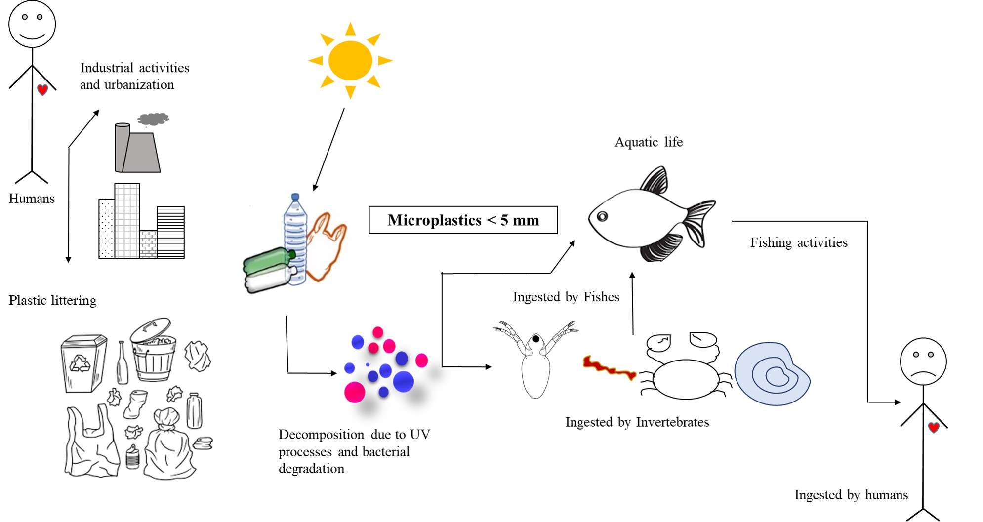
Figure 1. Incidence of microplastics in aquatic systems (adapted after Issac & Kandasubramanian, 2021)
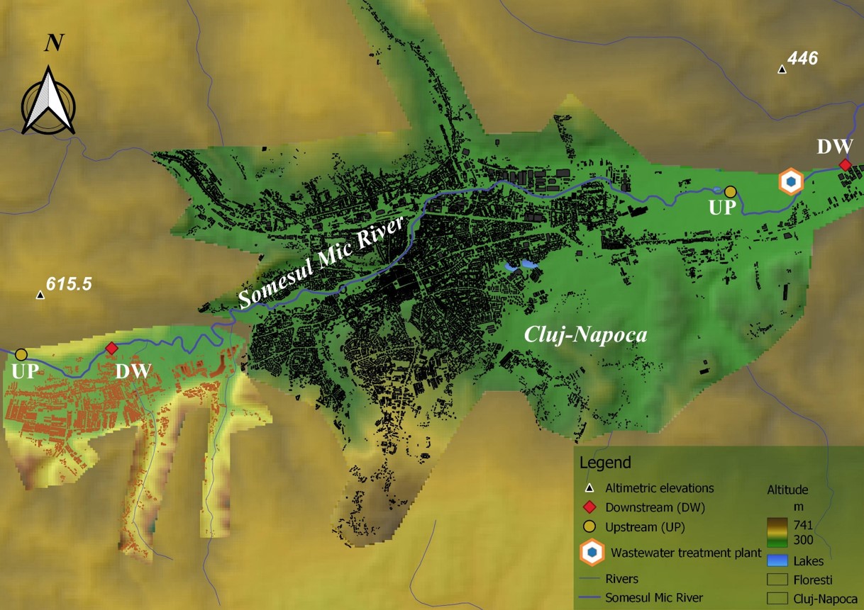
Figure 2. Sampling sites map
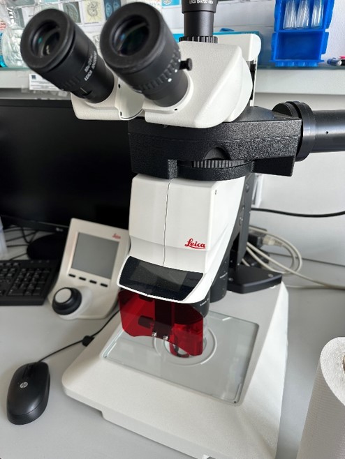
Figure 3.a. Visual microscopic analysis – Leica M205FA stereomicroscope
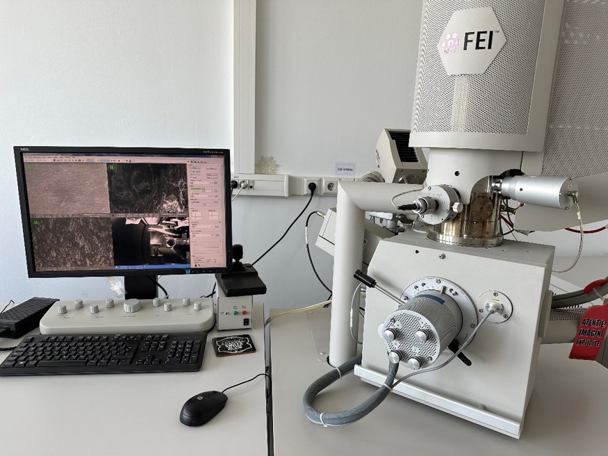
Figure 3.b. Visual microscopic analysis – SEM Quanta FEG 250
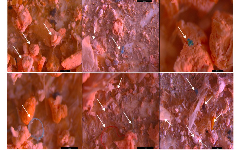
Figure 4. Stereomicroscopic analysis of microplastics – in the form of threads (red, blue, purple) and fragments (transparent, blue, green, yellow), foam spheres (white) collected on 200 µm filter, scale 1mm, 2mm, 500 µm, 1mm, 1mm, 2mm

Figure 5. SEM analysis of microplastics highlighted decomposed floating microplastics of spherical shape, scale 1 mm, 300 µm, 100 µm
